
New Wilf Campus Microscope Sheds Light on World of the Cell
Beginning this summer, Yeshiva University undergraduate researchers will be able to conduct cutting-edge scientific research using light microscopy, thanks to the generous donation of a confocal research microscope by Dr. Ruth Lehmann at the Skirball Institute for Biomolecular Medicine at New York University Langone Medical Center. This gift was coordinated by Dr. Josefa Steinhauer, assistant professor of biology at Yeshiva College.
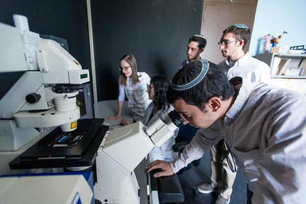 Students in the lab of Dr. Josefa Steinhauer use the confocal research microscope to study molecules within cells that have been labeled with light-sensitive chemicals.
“From the visualization of the first cells in 1665 by Robert Hooke, light microscopy has been at the core of biology,” said Steinhauer, who uses the microscope (manufactured by Carl Zeiss Inc) in her research on male fertility. “Today, light microscopy is used to visualize molecules within cells that have been labeled with light-sensitive chemicals called fluorophores, which fluoresce when illuminated. Confocal microscopy uses a focused beam of light—a laser—to illuminate the molecular labels inside cells, resulting in a crisp image that is free of background illumination. Since the 1990s, confocal microscopy has been the cornerstone of modern biological research.”
In Steinhauer’s lab, she and her students use fruit flies as a genetic model system to understand how sperm cells are formed, how diet influences fertility and how fertility declines with age. Because flies are evolutionarily related to humans, their studies have relevance for understanding human fertility in the context of our environment.
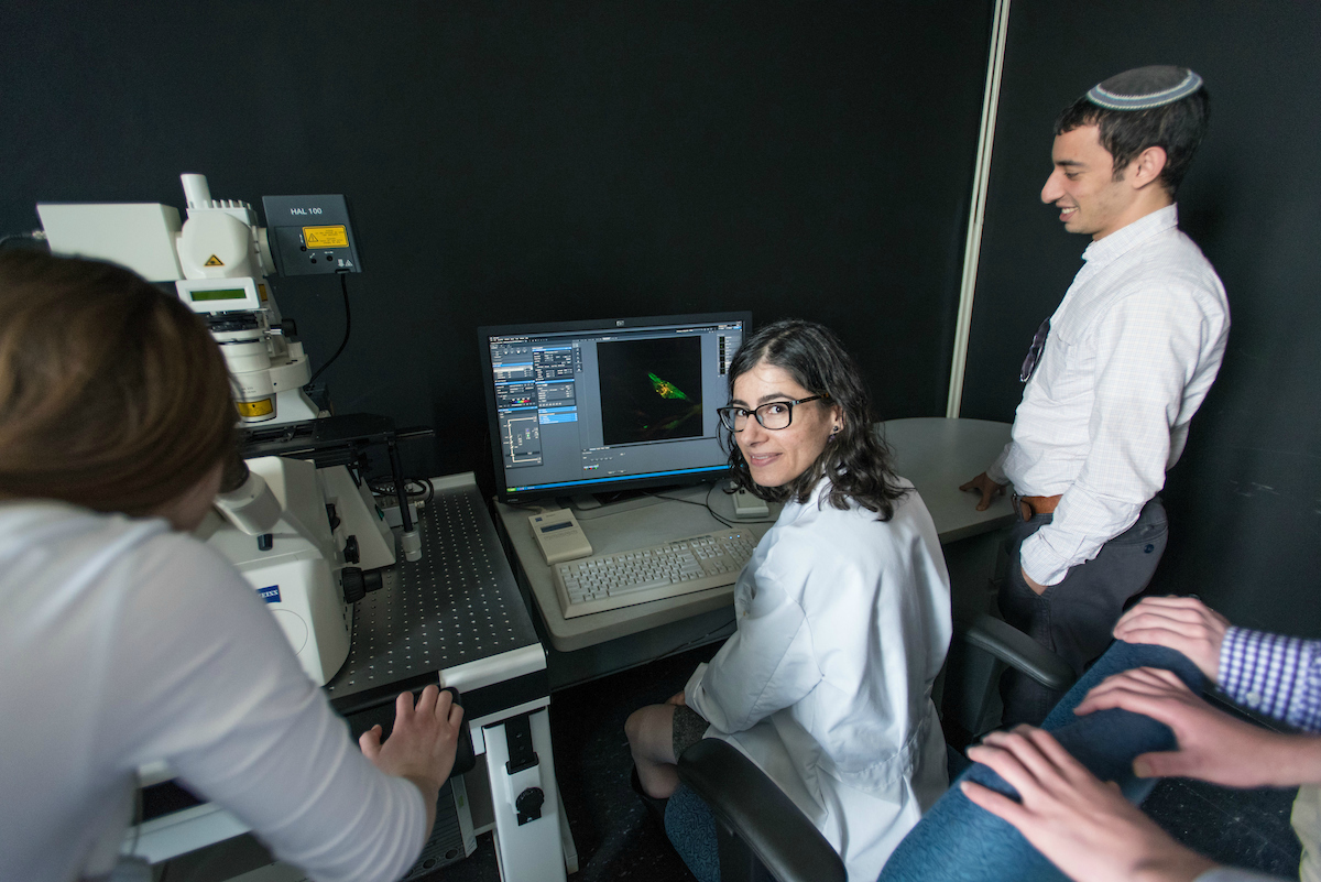 Steinhauer (center) uses light microscopy in her research on male fertility.
“This microscope allows the biology faculty here at the Wilf campus to further our research activities, which are a strong draw for our many pre-medical and science students,” said Steinhauer. “This equipment will enable us to produce publication-quality data and qualify for external funding from the National Institutes of Health, National Science Foundation and other granting agencies.”